

ZEISS Solutions for Geoscience
From Imaging to Analytics
Geoscience is a critical fundamental research topic focused on the examination of processes that govern the formation and evolution of the world around us. It also underpins the processes that control its economic development and utilization.
From micropaleontology to mineralogical studies to the modeling of three-dimensional fluid flow, ZEISS Microscopy has provided geoscientific imaging and analysis solutions for over one hundred years.
Discover the latest technical advancements:
Meet ZEISS at AGU 2023
11 - 15 December 2023
Visit us in Booth 523
Moscone Center in San Francisco, California, USA
Join us at the ZEISS Theatre for Expert Talks on
- Hard Rocks
- Soft Rocks
- Stratigraphy
- Planetary Geology
- Automated Petrology, Techniques and Analytics
- AI for Machine Learning, Segmentation, Analytics
- Paleontology and more...
Discover the Latest Technical Advancements


-


CHEMera mapping using ZEISS SEM with Mineralogic 2D combines geochemical variation with phase identification on Garnet-kyanite gneiss metamorphic rock from Glenelg, Scotland
ZEISS Mineralogic
Automated Quantitative Mineralogy
ZEISS Mineralogic 2D and 3D provide automated quantitative mineral analysis by bringing together cutting-edge microscopy with the scanning electron microscope (SEM) and the X-ray microscope (XRM), industry leading energy-dispersive spectroscopy (EDS), and AI-based deep learning algorithms to enhance your analytical capability and increase productivity.
ZEISS Mineralogic solutions are ideal for exacting geological interrogation of your samples, from in-depth petrological investigations to high throughput mineral liberation workflows to quantitative geochemistry.
Continue to swipe for more information →
-

Calcium heatmap highlights zoned garnet from Glenelg, Scotland. The intuitive periodic table user interface allows simple and dynamic visualization of geochemical data.
Mineralogic 2D
Benefits
- Make fact-based operational decisions with detailed mineral and sample chemical and particle information.
- Automatically categorize your particles, and identify and quantify ore types, with quantitative chemical analysis and classification.
- Combine chemistry with grain size, shape parameters, grey level, and porosity to achieve in-line morphochemical quantification with immediate results tabulation.
- Map samples for quantitative chemistry and combine with grain size, shape parameters, grey level, and porosity to achieve in-line morphochemical quantification with immediate results tabulation. Visualize and export geochemical data for flexible downstream workflows.
-

Exposing and classifying pyrite and chalcopyrite within gangue.
Mineralogic 3D
Benefits
- Investigate your sample in its true form, classifying mineralogy and measuring parameters in 3D, gaining an unparalleled ability to understand its composition, mineral relationships, and texture.
- Enjoy higher analysis throughput with simple sample handling by dispensing with the requirement to mechanically alter your samples in order to expose flat surfaces.
- Non-destructive imaging allows for analysis of precious samples or correlative workflows.


-

Automated Petrographic Analysis
Polarization microscopy faces a broad set of challenges when designing automated workflows to create digital petrographic records of slides. These unique challenges, broadly arising from the nature of the material under study, are overcome by the application of specialized equipment, automation, and computer vision techniques. The digital sections can then be studied with a virtual microscope, ideal for remote work and collaboration, and the depth of data generated is perfectly suited to downstream AI-integrated image analysis.

ZEISS Solutions for Geoscience ZEISS provides the most advanced solutions in 2D and 3D mineralogy, petrography, and mineral physics for your geoscience challenges, underscored by AI-driven segmentation and analysis..


Classification of shale heterogeneity. Green: fracture. Blue: low porosity. Red: high porosity. Yellow: pyrite. Imaged with ZEISS Xradia Versa 3D X-ray microscope.
Classification of shale heterogeneity. Green: fracture. Blue: low porosity. Red: high porosity. Yellow: pyrite. Imaged with ZEISS Xradia Versa 3D X-ray microscope.
Sedimentology
Perform detailed investigations of clastic, carbonate, and evaporitic rocks and understand weathering and erosion processes that shaped the geological features of Earth. Use automated grain size and shape measurements to understand the environmental conditions of formation. Employ correlative microscopy to blend mineralogy data from polarized light microscopes and automated mineralogy to provide textural knowledge. Determine stratigraphic sequences from microfossils and use detailed data on an organism’s structure to identify species and development levels to provide geological timescales.


Igneous & Metamorphic Petrology
Analyze and describe magmatic, volcanic, and metamorphic processes with advances in technology that allow you to quantify mineral distribution, automate large-scale analyses, describe structures in 3D, and integrate data together to facilitate a better understanding of the dynamic forces that shape the world around us.
Paleontology/Palynology
Traditional paleontological research has required samples to be removed from storage media or cut out from their host rock. Non-destructive imaging of irreplaceable samples can be accomplished using multiscale 3D X-ray microscopy. This allows you to make 3D morphological measurements on internal structures without interfering with the sample in any way.
Planetary Geology
From meteorites to pre-solar grains, the study of extra-terrestrial material requires advanced capabilities using multi-scale, multi-modal correlative analysis.


Volume segment showing interior location of gold in core sample
Volume segment showing interior location of gold in core sample
Ore Body Research
Improving ore deposit knowledge, refining our understanding of oregenesis and understanding more effective ways of extracting valuable ore is critical in ensuring we have resources available for future generations. For these studies we can combine optical, electron, and X-ray microscopy to help characterize and understand these ore deposits.


Mineral physics at the nanoscale with multidirectional magnetic fields


Mineral physics at the nanoscale with multidirectional magnetic fields. Image courtesy of University of Cambridge, UK.
Mineral physics at the nanoscale with multidirectional magnetic fields. Image courtesy of University of Cambridge, UK.
Mineral Physics
The science of mineral physics involves the understanding of the physical properties of the materials that comprise the Earth and other planets. It provides a critical link between fundamental petrological knowledge, and the more esoteric observations of seismic, magnetic, and geodynamic properties of rocks and the Earth. Understanding mineral properties at the micro-to-nano scale involves a combination of empirical observations of natural samples and experimental petrology. In other forms of geoscience, we look at a sample to understand something bigger; in mineral physics, we look at features much smaller than the sample.
Download this complimentary collection of papers
Applications and solutions for geoscience

-
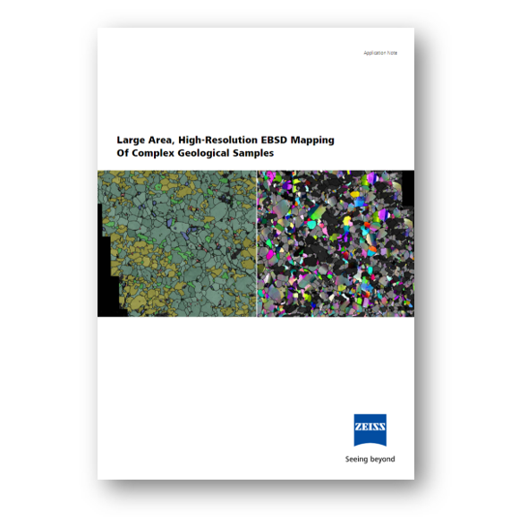
Large Area, High Resolution EBSD Mapping of Complex Geological Samples
Mapping the spatial distributions of crystalline phases and their lattice orientations using electron backscatter diffraction (EBSD) is a central task in both the materials and earth sciences, that also has application for extraterrestrial samples.
-
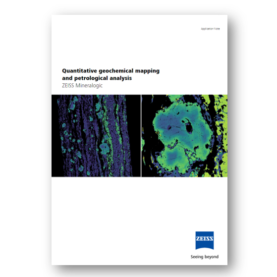
Quantitative geochemical mapping and petrological analysis
Achieve quantitative geochemical and petrological data of your extraterrestrial samples with the greatest possible flexibility.
-
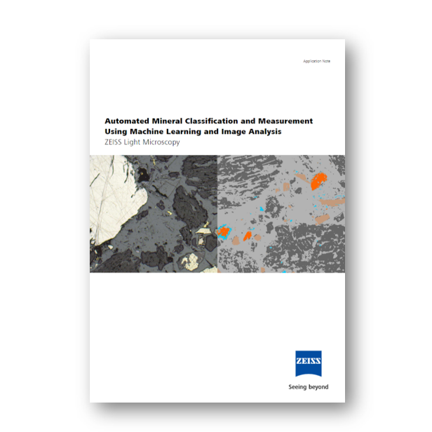
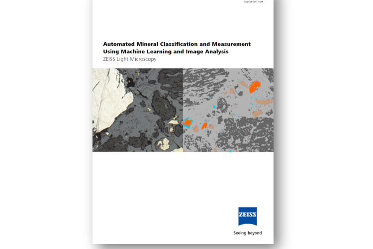
Automated Mineral Classification and Measurement Using Machine Learning and Image Analysis
ZEISS Light Microscopy
ZEN Intellesis plus ZEN Image Analysis modules offer machine learning image segmentation and classification, with image analysis and object measurements, for the light microscope. Available for use with the range of ZEISS light microscopes, from the operator-driven Axiolab 5 to the fully automated Axioscan 7 Geo, the modules offer automated identification, classification, measurement, and quantification of minerals.
-
Whole-particle Liberation Studies
ZEISS Mineralogic 3D
Mineralogic 3D – Automated Mineralogy
ZEISS Mineralogic 3D applies 3D X-ray microscopy techniques and deep learning algorithms to execute automated mineralogy analyses in three dimensions (fig 1) that provide particle identification, mineral classification, and data outputs including liberation and association measurements.Learn more about the three immediate benefits from analyzing in 3D.


From educating the next generation of geoscientists to the latest advances in technologies such as non-destructive 3D X-ray microscopy and quantitative mineral mapping, ZEISS enables you to gain unparalleled knowledge from your geoscientific specimens from the macro- to the nanoscale.


Download the Application Notes here ↓
After completion of the form all four application notes will be available for download


